Quick Answer for Stomach Ulcer vs Gastritis: Gastritis is diffuse inflammation restricted to the superficial gastric mucosa, causing a broad, gnawing epigastric ache and early satiety. A stomach ulcer is a focal structural defect penetrating deeper than 0.5 mm through the muscularis mucosae, causing sharp, localized burning and carrying a clinical risk of gastrointestinal bleeding.
Upper abdominal pain is notoriously deceptive. A patient with mild, superficial gastritis can experience severe, debilitating epigastric burning, while another patient with a deep, actively oozing duodenal ulcer may feel little more than vague afternoon hunger pangs until they develop iron-deficiency anemia. Because visceral pain receptors in the upper gastrointestinal tract lack the precise spatial mapping of skin receptors, the brain struggles to distinguish between diffuse surface irritation and a focal, deep tissue crater.
In clinical gastroenterology, separating gastritis from peptic ulcer disease (PUD) is the primary diagnostic priority when evaluating dyspepsia. While both conditions share common triggers—primarily Helicobacter pylori (H. pylori) infection, nonsteroidal anti-inflammatory drugs (NSAIDs), bile reflux, and physiological stress—their structural depth, complication profiles, and dietary tolerances differ substantially.
Gastritis is an inflammatory process confined to the innermost superficial layer of the stomach wall, the mucosa. A stomach ulcer (gastric ulcer) or duodenal ulcer is a structural excavation that penetrates completely through the mucosal layer, breaching the muscularis mucosae into the deeper submucosa or muscularis propria. That anatomical boundary—often less than a millimeter thick—is the dividing line between a condition managed with mucosal soothing and acid control, and one that carries a clinical risk of hemorrhage, perforation, or gastric outlet obstruction.
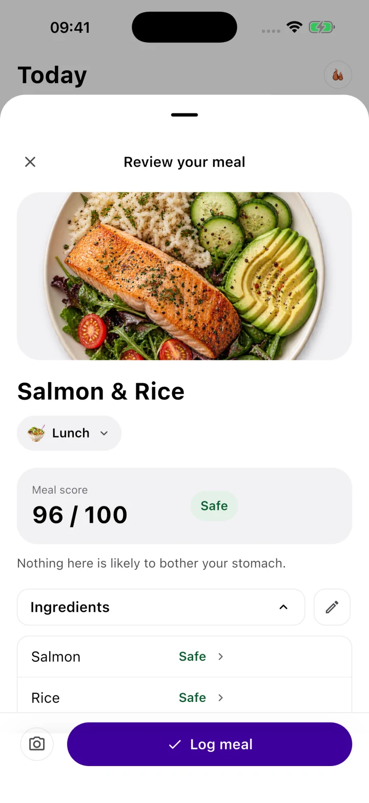
When every meal seems to provoke burning, pressure, or nausea, attempting to guess whether you are dealing with surface gastritis or an ulcer creates immense anxiety. Standard food diaries fail in this scenario because listing ingredients without accounting for their biomechanical and biochemical impact makes every food look like a culprit. In the Gutrack app, we replace guesswork with the Gastric Safety Index (GSI)—a 0 to 100 clinical scoring algorithm that evaluates meals based on titratable acidity, lipid transit delay, physical abrasiveness, and pepsinogen stimulation. By pairing GSI meal scores with precise postprandial symptom timestamps, patients and their gastroenterologists can observe how the stomach lining responds to specific physiological loads before and after an endoscopy.
Pathology & Histology: Surface Inflammation vs. Deep Excavation
To understand why an ulcer behaves differently than gastritis, we must examine the microscopic architecture of the gastric wall. From the inside of the stomach lumen outward, the wall is organized into four distinct concentric tunics:
- The Mucosa: Composed of the superficial simple columnar foveolar epithelium (which secretes protective mucus and bicarbonate), the lamina propria (loose connective tissue housing gastric glands, parietal cells, chief cells, and capillaries), and a thin lower boundary of smooth muscle called the muscularis mucosae.
- The Submucosa: A dense layer of connective tissue beneath the muscularis mucosae that contains larger arterioles, venules, lymphatics, and the Meissner’s (submucosal) nerve plexus.
- The Muscularis Propria: Three thick layers of smooth muscle (inner oblique, middle circular, outer longitudinal) responsible for peristaltic contractions and the mechanical grinding of food (antral trituration).
- The Serosa: The outermost fibrous membrane covering the stomach inside the peritoneal cavity.
How the Mucosal Defense Barrier Works
Under healthy conditions, the stomach lumen maintains a highly corrosive pH between 1.5 and 2.5, driven by hydrochloric acid ($\text{HCl}$) secreted by parietal cells. Simultaneously, chief cells release pepsinogen, an inactive zymogen that rapidly cleaves into active pepsin whenever the local pH drops below 4.0. Pepsin is a potent proteolytic enzyme designed to digest dietary collagen and muscle fibers.
The stomach avoids digesting itself through a multi-tiered defense system:
- Pre-epithelial defense: A continuous, water-insoluble viscoelastic gel layer of MUC5AC and MUC6 mucins secreted by surface foveolar cells and mucous neck cells. Bicarbonate ions ($\text{HCO}_3^-$) are actively pumped into this gel layer, establishing a pH gradient. Even when the lumen is at pH 1.5, the immediate surface of the epithelial cells remains buffered at a neutral pH 6.5 to 7.0, keeping pepsin completely inactivated at the cellular border.
- Epithelial defense: Intercellular tight junctions prevent acid back-diffusion between cells. Furthermore, if surface cells are damaged, adjacent healthy cells rapidly migrate across the basal lamina to seal the gap within hours—a process called mucosal restitution.
- Post-epithelial defense: Dense microvascular blood flow in the lamina propria supplies oxygen and nutrients while rapidly washing away any hydrogen ions ($H^+$) that manage to diffuse through the mucus layer.
The Histological Boundary: Gastritis vs. Erosion vs. Ulcer
When noxious agents overwhelm these defenses, injury progresses along a strict pathological spectrum:
- Non-Erosive Gastritis: Microscopic infiltration of inflammatory cells (neutrophils in acute or active chronic phases; lymphocytes and plasma cells in chronic phases) within the lamina propria, accompanied by foveolar hyperplasia (reactive thickening of surface cells) or mucosal edema. The epithelial surface remains continuous and intact.
- Erosive Gastritis (Gastric Erosions): Shallow breaks in the superficial epithelium that extend into the lamina propria but do not cross the muscularis mucosae. Because large blood vessels do not reside in the superficial lamina propria, erosions may ooze tiny amounts of capillary blood, but they rarely cause life-threatening hemorrhage and heal without leaving fibrous scar tissue.
- Peptic Ulcer: A deep, well-circumscribed mucosal break measuring at least 5 mm (0.5 cm) in diameter that penetrates completely through the muscularis mucosae into the submucosa or deeper. Histologically, a chronic peptic ulcer crater exhibits four classic zones (Askanazy zones): a superficial layer of fibrinopurulent exudate and necrotic debris, a zone of fibrinoid necrosis, a deeper layer of vascular granulation tissue, and a dense base of collagenous fibrous scar tissue. Because the ulcer crater breaches the submucosa, it directly exposes the larger arterioles of the submucosal plexus and unmyelinated pain fibers to gastric acid and pepsin.
Primary Biochemical Drivers of Deep Excavation
1. NSAIDs and COX-1 Prostaglandin Depletion
Nonsteroidal anti-inflammatory drugs (ibuprofen, naproxen, aspirin, ketorolac, meloxicam) damage the stomach through two distinct mechanisms: topical ion trapping and systemic cyclooxygenase-1 (COX-1) inhibition.
Most NSAIDs are weak organic acids. In the highly acidic gastric juice (pH 2.0), they remain non-ionized and lipid-soluble, allowing them to freely diffuse across the lipid bilayer membranes of surface epithelial cells. Once inside the cell—where the cytoplasmic pH is neutral (pH 7.0)—the NSAID molecule dissociates into an ionized state. This ion trapping concentrates the drug inside the epithelial cell at levels vastly higher than in the lumen, uncoupling oxidative phosphorylation, depleting cellular ATP, and increasing cell membrane permeability to acid and bile salts.
More critically, NSAIDs inhibit the COX-1 enzyme, blocking the conversion of arachidonic acid into endogenous prostaglandins ($\text{PGE}_2$ and $\text{PGI}_2$). Prostaglandins are the master regulators of gastric cytoprotection: they stimulate mucin and bicarbonate secretion, inhibit parietal cell acid output, and maintain vasodilatory blood flow in the mucosal microcirculation. Even if an NSAID is taken intravenously or via suppository, systemic COX-1 inhibition strips away the mucus-bicarbonate shield and induces microvascular ischemia, allowing luminal acid and pepsin to excavate an ulcer crater.
2. Helicobacter pylori Virulence Factors
H. pylori is a spiral, flagellated Gram-negative bacterium uniquely adapted to survive in the stomach. It secretes large quantities of the enzyme urease, which hydrolyzes luminal urea into ammonia ($\text{NH}_3$) and carbon dioxide, creating a localized alkaline cloud around the bacterium. Using its flagella, H. pylori burrows through the viscoelastic mucus layer to attach directly to surface foveolar cells.
Strains expressing the CagA (cytotoxin-associated gene A) and VacA (vacuolating cytotoxin A) proteins inject bacterial effectors directly into host epithelial cells, disrupting tight junctions and triggering an intense inflammatory cascade (interleukin-8 release) that recruits neutrophils. Over time, depending on where the infection settles, it drives two different ulcer pathways:
- Antral-predominant gastritis (Duodenal Ulcer phenotype): Inflammation localized to the gastric antrum destroys somatostatin-producing D-cells. Without somatostatin to apply the brakes on G-cells, gastrin secretion surges (hypergastrinemia). This drives the healthy parietal cells in the stomach body to hypersecrete massive amounts of hydrochloric acid. When this hyperacidic chyme dumps into the duodenal bulb, the duodenum undergoes protective gastric metaplasia, which H. pylori then colonizes and ulcerates.
- Pangastritis / Corpus-predominant gastritis (Gastric Ulcer phenotype): Inflammation spreads across the entire stomach body, damaging parietal cells. Acid output here is often normal or even low (hypochlorhydria), but the mucosal barrier is so severely degraded by chronic inflammation and atrophy that even modest amounts of acid and pepsin excavate a gastric ulcer—most commonly along the lesser curvature at the incisura angularis, where blood supply is anatomically tenuous and mechanical shear stress is high.
Symptom Chronology: Decoding Pain Timing After Meals
While an endoscopy is required for definitive structural diagnosis, a careful clinical history focusing on postprandial symptom chronology—exactly how many minutes or hours after eating pain begins or subsides—provides strong diagnostic differentiation between gastritis, a gastric ulcer, and a duodenal ulcer.
| Clinical Feature | Chronic / Erosive Gastritis | Gastric Ulcer (Stomach) | Duodenal Ulcer (First Part of Small Intestine) |
|---|---|---|---|
| Anatomical Depth | Confined to superficial mucosa; does not breach muscularis mucosae. | Penetrates through muscularis mucosae (>0.5 mm depth) in the stomach (often lesser curvature). | Penetrates through muscularis mucosae in the duodenal bulb (first 2–3 cm past pylorus). |
| Pain Character & Location | Diffuse, dull burning, heavy pressure, or aching across the entire upper abdomen (epigastrium). | Sharp, boring, focal burning pain; patient can often point to a single spot in the midline or left epigastrium. | Deep, gnawing, "acid-hunger" burning localized slightly to the right of the midline epigastrium. |
| Immediate Meal Response (0–30 Mins) | Variable; large volume or acidic/fatty meals trigger immediate bloating, fullness, and burning. | Worsens rapidly with food. Eating stimulates gastrin, acid, and antral grinding directly over the raw crater. | Temporarily relieved by food. Food buffers acid, and the pyloric sphincter closes to retain food in the stomach. |
| Delayed Meal Response (2–4 Hours) | Gradually settles as the stomach empties, unless high-fat meals delay gastric emptying via CCK. | Pain subsides gradually as the stomach empties and meal-stimulated acid secretion declines. | Pain returns intensely 2–3 hours after eating as buffered food leaves and unbuffered acid dumps into the duodenum. |
| Nocturnal Pain (1:00 AM – 3:00 AM) | Less common unless accompanied by nocturnal gastroesophageal reflux (GERD) or bile reflux. | Occasional, though less predictable than duodenal ulcers. | Classic hallmark. Circadian histamine and vagal peaks drive unbuffered acid into the empty duodenum, waking the patient. |
| Impact on Appetite & Weight | Early satiety (feeling full after 4–5 bites), mild nausea, cautious eating. | Food aversion and weight loss. Patients fear eating because meals reliably trigger sharp pain. | Weight maintenance or slight weight gain; patients frequently snack to buffer the gnawing pain. |
1. The Gastric Ulcer Timeline: Immediate Postprandial Aggravation
In a patient with an active gastric ulcer, the protective mucus layer is completely absent over the crater base, leaving raw granulation tissue and submucosal nerve endings directly exposed to the stomach lumen.
When the patient begins eating, three physiological events occur within 5 to 30 minutes:
- Cephalic and Gastric Phase Acid Surge: The sight, smell, and physical distension of the stomach wall activate the vagus nerve (releasing acetylcholine) and antral G-cells (releasing gastrin). Parietal cells immediately pump $\text{HCl}$ into the lumen, while chief cells release pepsinogen.
- Mechanical Antral Trituration: To break solid food down into particles smaller than 2 mm so they can pass through the pylorus, the distal stomach initiates forceful peristaltic waves (3 contractions per minute). This physically grinds food particles directly across the exposed ulcer bed.
- Isometric Wall Tension: Stretching the inflamed muscularis propria pulls on the fibrous base of the ulcer crater.
Consequently, patients with a gastric ulcer frequently report that pain strikes within 15 to 30 minutes of starting a meal. Because eating reliably hurts, patients develop conditioned food aversion, skip meals, and often present to their gastroenterologist with unintended weight loss.
2. The Duodenal Ulcer Timeline: The "Buffer and Burn" Cycle
Duodenal ulcers—which account for roughly 70% to 80% of all peptic ulcers in younger populations—follow the opposite chronology. These ulcers almost always reside within the first 2 to 3 centimeters of the duodenal bulb, immediately downstream from the pyloric sphincter.
When a patient with a duodenal ulcer is fasting, basal acid secreted by the stomach trickles continuously through the relaxed pylorus into the duodenum, bathing the ulcer crater in unbuffered acid and causing a persistent, gnawing "hunger pain."
When the patient eats a meal:
- Immediate Buffering & Pyloric Closure: Proteins and carbohydrates in the meal temporarily absorb free hydrogen ions in the stomach, raising intragastric pH from ~1.5 up toward 4.0–5.0. Simultaneously, the pyloric sphincter tightens its tone to hold solid food in the stomach for mechanical grinding, temporarily stopping acid from entering the duodenum. Meanwhile, the arrival of nutrients stimulates duodenal S-cells to release secretin, which prompts the pancreas and Brunner’s glands to flood the duodenal bulb with alkaline bicarbonate. Result: The pain vanishes within minutes of eating.
- The 2-to-3 Hour Rebound Burn: By 120 to 180 minutes after the meal, the buffered food chyme has mostly emptied past the duodenum into the jejunum. However, parietal cells remain in a state of high meal-stimulated acid secretion. Now, pure, unbuffered hydrochloric acid and active pepsin pour through the pylorus directly onto the raw duodenal crater. Result: Deep burning pain returns sharply 2 to 3 hours after eating.
- The 2:00 AM Circadian Awakening: Basal gastric acid secretion follows a circadian rhythm driven by vagal tone and histamine release from enterochromaffin-like (ECL) cells, peaking between 11:00 PM and 2:00 AM. Because there is no food in the stomach at night to buffer this surge, patients with duodenal ulcers are classically awakened from sleep between 1:00 AM and 3:00 AM with sharp epigastric burning that improves if they drink milk, take an antacid, or eat a bland cracker.
3. The Gastritis Timeline: Diffuse Volume and Chemical Sensitivity
In chronic or erosive gastritis, the pain is rarely as clockwork-precise as a duodenal ulcer. Because inflammation is spread across the antrum or corpus rather than concentrated in a single deep crater, symptoms are governed by intragastric volume, chemical osmolarity, and transit speed:
- Early Satiety and Epigastric Pressure: Chronic mucosal inflammation impairs gastric accommodation—the vagally mediated reflex where the upper stomach (fundus) relaxes to expand and hold a meal without increasing internal pressure. In gastritis, the fundus remains stiff; eating even a small 250-gram meal increases intragastric pressure, firing stretch mechanoreceptors that the brain interprets as heavy fullness, bloating, and upper abdominal ache.
- Chemical Nociception: Because tight junctions between surface cells are leaky, acidic foods (pH < 4.0), alcohol, black pepper (piperine), chili (capsaicin, which binds TRPV1 pain receptors), and concentrated simple sugars diffuse rapidly into the lamina propria, causing an immediate, wide burning sensation across the upper abdomen within minutes of ingestion.
Red Flag Symptoms: Recognizing Upper GI Bleeding and Complications
Because a peptic ulcer penetrates into the submucosa—and occasionally deeper into the muscularis propria or through the serosa—it sits in direct anatomical proximity to substantial arterial blood vessels. Specifically, ulcers along the posterior wall of the duodenal bulb sit directly over the gastroduodenal artery, while ulcers along the lesser curvature of the stomach sit near branches of the left gastric artery.
If pepsin and acid erode through the fibrous wall of an arteriole at the base of an ulcer crater, upper gastrointestinal bleeding (UGIB) occurs. Every patient managing gastritis or suspected ulcer symptoms must know how to recognize both slow microvascular oozing and acute arterial bleeding.
1. Melena (Black, Tarry Stools)
When blood enters the stomach or duodenum, hydrochloric acid and digestive enzymes oxidize the iron inside hemoglobin molecules, converting red hemoglobin into hematin, a jet-black pigment. As this altered blood travels through the small intestine and colon (typically taking 8 to 14 hours), bacterial enzymes further degrade it.
- What it looks like: Stools that are jet-black (like road tar), sticky, glistening, and carry a distinct, metallic, foul odor. It takes as little as 50 to 100 mL of blood in the upper GI tract to produce a single melenic stool.
- Important Clinical Caveat: Always check your medication list before panicking over black stool. Bismuth subsalicylate (Pepto-Bismol) reacts with trace sulfur in saliva and the GI tract to form bismuth sulfide, which turns both the tongue and stool completely black. Similarly, oral iron supplements (ferrous sulfate), activated charcoal, and massive servings of dark blueberries or black licorice can mimic dark stool, though they do not produce the sticky, tarry consistency or foul smell of true melena. If you are not taking bismuth or iron, black tarry stool requires urgent medical evaluation within hours.
2. Hematemesis (Vomiting Blood or "Coffee Grounds")
If an ulcer bleeds more briskly inside the stomach, blood accumulates in the lumen, irritating the gastric wall and triggering the vomiting reflex:
- Coffee-Ground Emesis: When blood sits in the stomach for 15 to 60 minutes before being vomited, gastric acid clots and oxidizes the blood into dark brown or black granular particles that look identical to wet coffee grounds. This indicates recent or ongoing upper GI bleeding and is a medical emergency.
- Bright Red Hematemesis: Vomiting fresh, bright red blood or maroon clots indicates brisk, active arterial bleeding that is occurring too rapidly for stomach acid to oxidize it. Call emergency services immediately.
3. Occult Bleeding and Hemodynamic Shifts
Many gastric and duodenal ulcers never cause dramatic vomiting or obvious melena; instead, they ooze 5 to 15 mL of blood per day invisibly into the stool (fecal occult blood). Over weeks, this depletes the body’s iron stores (ferritin), preventing bone marrow from synthesizing adequate hemoglobin. Watch for:
- Orthostatic Dizziness (Postural Hypotension): Feeling lightheaded, seeing dark spots, or feeling your heart race (tachycardia) within 60 seconds of standing up from a lying or seated position.
- Exertional Dyspnea and Fatigue: Becoming unusually short of breath while walking up a single flight of stairs, accompanied by pale conjunctivae (the inner lower eyelids looking pale pink instead of vibrant red).
4. Perforation and Penetration
If an ulcer erodes completely through all four layers of the stomach or duodenal wall (free perforation), acidic gastric contents spill directly into the sterile peritoneal cavity, causing chemical and bacterial peritonitis.
- Symptom: Sudden, catastrophic, knife-like upper abdominal pain (thunderclap abdominal pain) where the patient can pinpoint the exact minute the pain started. The abdominal muscles involuntarily contract into a rigid, board-like wall (peritoneal guarding), and any movement, coughing, or deep breathing severely intensifies the pain. This requires emergency surgical repair.
5. Gastric Outlet Obstruction (Pyloric Stenosis)
When an ulcer is located inside the pyloric channel or duodenal bulb, repeated cycles of acute inflammation (mucosal edema and spasm) or chronic healing (collagenous fibrotic scarring) can narrow the exit passage of the stomach.
- Symptom: Progressive bloating, persistent nausea, loud sloshing sounds in the upper abdomen hours after eating (succussion splash), and eventually vomiting large volumes of undigested food eaten 6 to 12 hours earlier.
Why Standard Food Diaries Fail in Ulcer & Gastritis Care
When patients experience severe epigastric pain, their natural instinct is to start a notes app or paper food diary. Within five days, almost every patient reaches the same frustrating conclusion: "Everything I eat hurts, even plain water."
From a physiological standpoint, unstructured food logging fails for three reasons:
- Delayed Transit Masking: If a patient eats a high-fat meal at 12:30 PM (such as salmon with avocado oil), duodenal I-cells release cholecystokinin (CCK), which slows gastric emptying and relaxes the lower esophageal sphincter. That meal may sit in the stomach for 4 to 5 hours. When the patient drinks a glass of plain water or eats a banana at 4:00 PM, the mechanical volume stretch on top of the unemptied lunch triggers severe pain. A basic food log blames the water or banana at 4:00 PM, completely missing the CCK-mediated lipid delay from 12:30 PM.
- Hidden Biochemical Triggers: Logging "chicken soup" tells a clinician nothing. Was it made with onion and garlic powder (high fructans and transient LES relaxants), black pepper (mucosal irritant), and lemon juice (pH 2.3, instantly activating pepsin)? Or was it pure collagen bone broth with white rice and skinless chicken breast (pH 6.2, low fat, high GSI)?
- Lack of Chronological Windowing: As demonstrated in our clinical comparison table, pain occurring 20 minutes after a meal points toward gastric mucosal irritation or a gastric ulcer, whereas pain occurring 150 minutes after a meal points toward duodenal acid exposure. Standard trackers do not correlate symptom onset against the gastric emptying curve.
This is why we built Gutrack. When you scan or log a meal in Gutrack, the app calculates an instant 0 to 100 Gastric Safety Index (GSI) score before you take a bite, breaking down acidity, fat load, fiber roughness, and spice irritants. When you log a symptom—such as focal epigastric burning or early satiety—Gutrack automatically maps the exact postprandial time elapsed since your last meal and medication dose, compiling these patterns into a clean 14-day Clinical PDF Report designed specifically for your gastroenterology consultation.
The GSI Cytoprotective Nutrition Matrix for Ulcers & Gastritis
Whether you are waiting for an upper endoscopy or actively healing a confirmed peptic ulcer alongside proton pump inhibitors (PPIs) and sucralfate, your dietary objective is mechanical and chemical cytoprotection.
Every meal must achieve four physiological goals:
- Maintain Intragastric pH > 4.0: Avoid introducing dietary acids (citric, malic, acetic, ascorbic, or chlorogenic acids) that drop luminal pH below 4.0, where inactive pepsinogen cleaves into tissue-digesting pepsin.
- Minimize Antral Grinding (Trituration): Select foods that are already soft, emulsified, or easily broken down into particles <2 mm so the distal stomach does not have to perform forceful peristaltic grinding across an inflamed or ulcerated mucosa.
- Accelerate Gastric Emptying (Low CCK Load): Keep single-meal lipid content below 8 to 10 grams of fat. Dietary fat triggers duodenal cholecystokinin (CCK) release, which inhibits antral motility and traps acidic chyme inside the stomach for hours.
- Provide Mucilaginous and Amino Acid Substrates: Incorporate soluble beta-glucans (which form a physical hydrocolloid gel over the mucosa) and high-bioavailability proteins rich in L-glutamine, glycine, and proline—the primary amino acids required by epithelial cells for tight-junction synthesis and collagen repair in the ulcer bed.
Below is the Gastric Safety Index (GSI) Cytoprotective Matrix, contrasting therapeutic ulcer-safe meals against common traps that delay mucosal healing.
| Meal / Food Item | GSI Score (0–100) | GSI Safety Zone | Physiological & Biochemical Mechanism on Ulcer / Gastritis Mucosa |
|---|---|---|---|
| Slow-Simmered White Rice Congee in Skimmed Collagen Bone Broth | 96 / 100 | Safe (70–100) | Extended cooking gelatinizes starch completely, requiring zero mechanical antral grinding. Skimmed broth (0g fat) delivers free glycine, proline, and glutamine to support mucosal restitution at a neutral pH (~6.3) with rapid gastric emptying (<90 mins). |
| Steamed Atlantic Cod (120g) with Pureed Butternut Squash | 93 / 100 | Safe (70–100) | Cod contains <1g lipid per 100g with extremely short, fragile muscle fibers (minimal myomesin connective tissue) that dissolve rapidly in the stomach while buffering free H+ ions. Squash provides provitamin A carotenoids for epithelial differentiation without insoluble roughage. |
| Poached or Soft-Scrambled Egg Whites with Rolled Oat Porridge (Water/Almond Milk) | 91 / 100 | Safe (70–100) | Removing the egg yolk eliminates ~5g of saturated/monounsaturated fat per egg (avoiding CCK transit delay) while preserving pure ovalbumin protein to buffer luminal acid. Oat beta-glucans form a soothing, viscous colloidal film over raw foveolar cells. |
| Silken Tofu & Wakame Miso Broth (Low-Sodium, Lukewarm) | 86 / 100 | Safe (70–100) | Silken tofu is coagulated soy protein with a high water content and soft curds that pass through the pylorus effortlessly. Wakame seaweed provides sodium alginate, which reacts with gastric acid to form a protective floating raft. Served at <50°C to avoid thermal hyperemia. |
| Whole-Wheat Toast with 2 Tbsp Creamy Peanut Butter & Sliced Banana | 54 / 100 | Caution (40–69) | While banana is alkaline and soothing (contains leucocyanidin, which promotes mucus thickness), 2 tablespoons of peanut butter delivers 16g of dense lipid, triggering strong CCK release and delaying gastric emptying by 2+ hours. Whole-wheat bran shards can mechanically abrade active erosions. |
| Greek Yogurt Bowl with Fresh Strawberries, Blueberries & Granola | 42 / 100 | Caution (40–69) | A classic "healthy" trap for active ulcers. Strawberries and blueberries have a titratable pH between 3.1 and 3.5 (citric and malic acid), instantly activating surface-bound pepsin on the ulcer crater. Baked oat/nut granola clusters act as rigid mechanical abrasives during antral trituration. |
| Decaf Black Coffee or Citrus Herbal Tea on an Empty Stomach | 24 / 100 | High Risk (0–39) | Even without caffeine, roasted coffee beans contain rich concentrations of chlorogenic acids and N-alkanoyl-5-hydroxytryptamides (C5HTs) that directly stimulate parietal cell H+/K+-ATPase proton pumps. Citrus hibiscus tea carries a pH of ~2.6, reactivating dormant mucosal pepsin. |
| Pan-Seared Ribeye Steak with Garlic-Herb Butter & Red Wine | 08 / 100 | High Risk (0–39) | Dense mammalian elastin and collagen fibers require hours of intense acid-pepsin digestion and forceful antral grinding. High saturated fat (35g+) halts gastric emptying via CCK, while ethanol (12–14% ABV) dissolves the superficial MUC5AC mucus gel and causes direct microvascular congestion. |
Deep Dive: Three Therapeutic Meals for an Active Ulcer or Flare
1. The Morning Buffer: Oat Beta-Glucan & Egg White Custard Porridge (GSI: 91)
During the early morning hours, residual nocturnal acid sits in the empty stomach. Waiting until noon to eat ("intermittent fasting") is destructive for both duodenal ulcers and chronic gastritis because unbuffered acid and bile salts bathe the bare mucosa for hours.
- Clinical Preparation: Simmer 40g of fine rolled oats in 250 mL of water or unsweetened fortified almond milk (pH ~7.8 due to added calcium carbonate, which acts as a natural antacid buffer) for 8 minutes until completely soft. Reduce heat to low and slowly whisk in 3 liquid egg whites (approx. 100g) for 2 minutes until silky and cooked through.
- Why it works: The soluble $\beta$-(1,3)(1,4)-D-glucan polysaccharides leach out of the oat endosperm to create a thick mucilaginous hydrocolloid that coats the stomach lining, while the pure egg albumin binds free hydrogen ions without introducing egg yolk lipids.
2. The Midday Restitution Meal: Skimmed Bone Broth Rice Congee (GSI: 96)
Traditional Asian rice congee (jook) is arguably the highest-scoring meal in clinical gastroenterology for an acute ulcer flare.
- Clinical Preparation: Combine 1 part white jasmine or short-grain sushi rice with 8 parts fat-skimmed chicken bone broth (ensure all solidified yellow fat has been chilled and scraped off the surface; zero onion or garlic in the broth). Simmer on low heat for 60 to 90 minutes, stirring occasionally, until the individual rice grains rupture into a smooth, creamy porridge. Season lightly with a pinch of sea salt (avoid excessive hypertonic sodium, which irritates gastric mucosa).
- Why it works: White rice has had its insoluble fibrous bran hull removed. Boiling it in an 8:1 liquid ratio completely hydrolyzes the amylopectin starch matrix, meaning salivary and pancreatic amylases can digest it almost instantly without requiring mechanical grinding by the stomach antrum. Meanwhile, the collagen peptides supply 10 to 15 grams of pure protein per bowl, rich in L-glutamine—the primary metabolic fuel source for rapidly dividing gastrointestinal enterocytes.
3. Thermal and Osmotic Rules During Ulcer Healing
Beyond macro-ingredients, how you consume food alters mucosal blood flow and irritation:
- Thermal Neutrality (37°C to 50°C): Piping hot soups or teas (>60°C / 140°F) cause direct thermal injury and reactive vasodilation (hyperemia) in the fragile granulation tissue of an ulcer bed, increasing the risk of capillary oozing. Conversely, ice-cold drinks slow enzymatic activity and can trigger pyloric spasm. Aim for lukewarm to warm temperatures.
- Meal Volume Cap (<350 mL / grams per sitting): Eat 5 small meals spaced every 3 hours rather than 2 or 3 large meals. Keeping total meal volume under 350 mL prevents mechanical stretch of the stomach wall, keeping gastrin release mild while maintaining a continuous, gentle protein buffer inside the lumen to protect duodenal ulcers.
Preparing for Your Upper Endoscopy (EGD): A Patient Guide
If you have persistent epigastric pain lasting more than two weeks, incomplete relief from acid suppression, or any red flag symptoms, your gastroenterologist will schedule an Esophagogastroduodenoscopy (EGD)—commonly called an upper endoscopy.
While an H. pylori urea breath test or stool antigen test
Clinical Evidence & Scientific References
- Laine L, et al. Management of Patients with Ulcer Bleeding: An Updated ACG Guideline. Am J Gastroenterol. 2021;116(5):899-917.
- Malfertheiner P, et al. Management of Helicobacter pylori infection: the Maastricht VI/Florence consensus report. Gut. 2022;71(9):1724-1762.
- National Institute of Diabetes and Digestive and Kidney Diseases (NIDDK). Peptic Ulcer Disease: Diagnosis, Symptoms, and Bleeding Complications. NIH Pub. No. 22-5140.
Self-tracking tool, not clinical diagnosis
Gutrack provides evidence-based Gastric Safety Index estimates for daily self-management and diet optimization. It does not replace medical consultation, endoscopy, or treatment prescribed by a qualified gastroenterologist.
Frequently asked questions
How do you know if you have an ulcer or just gastritis?
Symptom patterns provide strong clinical clues, such as localized sharp pain triggered within 30 minutes of eating for gastric ulcers versus diffuse upper abdominal pressure for gastritis. However, an upper endoscopy (EGD) is the only definitive way to visually confirm whether the lesion breaches the muscularis mucosae (>5 mm diameter).
What does ulcer pain feel like compared to gastritis burning?
Gastritis typically presents as a dull, diffuse burning or heavy fullness across the entire upper abdomen that fluctuates with meal volume and acidity. Peptic ulcer pain is sharper, deeply boring, and localized to a specific finger-point spot in the epigastrium, often waking patients between 1:00 AM and 3:00 AM during circadian acid peaks.
What are the early warning signs of internal gastric bleeding?
Slow upper gastrointestinal bleeding presents as melena (jet-black, tarry, foul-smelling stools caused by acid oxidizing hemoglobin into hematin) and unexplained iron-deficiency anemia or orthostatic dizziness. Acute bleeding causes hematemesis—vomiting bright red blood or dark 'coffee-ground' material—requiring immediate emergency endoscopic intervention.
Can chronic gastritis turn into a peptic ulcer if left untreated?
Yes, untreated chronic gastritis compromises the mucus-bicarbonate barrier, allowing hydrochloric acid and activated pepsin (active at pH < 4.0) to digest the underlying epithelial cells. In the presence of Helicobacter pylori or daily NSAID use, superficial erosions can progress into full-thickness peptic ulcers within weeks to months.
This article is for educational purposes only and isn't a substitute for professional medical advice, diagnosis, or treatment. Gutrack is a self-tracking tool, not a medical device. If you're experiencing persistent or concerning symptoms, talk to a doctor.